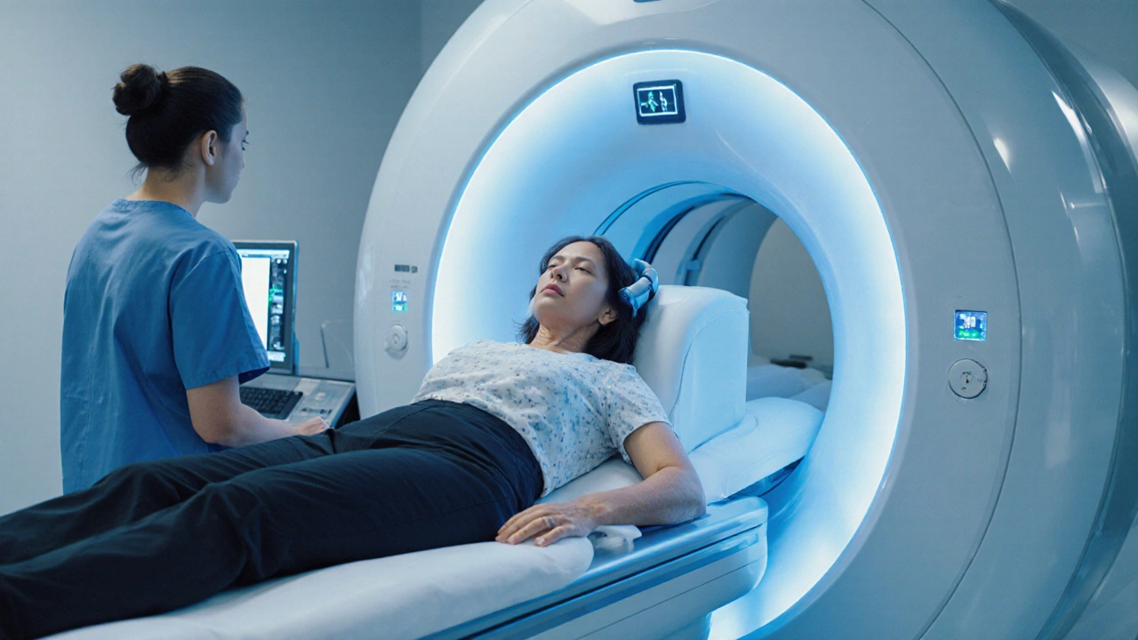
Explore how MRI works to diagnose Clinically Isolated Syndrome, the key imaging protocols, typical findings, and next steps for patients and clinicians.
MRI: Magnetic Resonance Imaging Explained
When working with MRI – magnetic resonance imaging, a non‑invasive technique that uses strong magnetic fields and radio waves to generate detailed internal images of the body. Also called magnetic resonance scan, it helps doctors diagnose conditions without ionizing radiation. In the world of medical imaging, MRI stands out because it can visualize soft tissues—like brain, muscle, and ligaments—much clearer than X‑ray or CT. The technology relies on three core components: a powerful magnet that aligns hydrogen protons, a radiofrequency pulse that nudges those protons, and a computer that translates the resulting signals into pictures. This process produces cross‑sectional slices that can be stacked into 3‑D models, allowing physicians to spot tumors, tears, or inflammation early. Safety‑wise, the lack of radiation makes MRI a go‑to option for pregnant patients and children, though the strong magnetic field means metal implants must be screened. Understanding the physics behind the scan helps patients feel less anxious and clinicians to choose the right protocol for each case.
Advanced MRI Techniques and Related Fields
Beyond the basic scan, several specialized techniques expand what MRI can reveal. functional MRI – a brain‑focused method that tracks blood‑oxygen‑level changes to map neural activity turns the machine into a window on thought processes, helping surgeons avoid critical areas before operations and researchers study memory or language. Another important addition is gadolinium‑based contrast agents – injectable substances that shorten signal relaxation times, making blood vessels and lesions stand out sharply on images. When used wisely, contrast enhances the detection of small tumors or vascular malformations that might blend into surrounding tissue on a plain scan. The broader field that houses all these tools is radiology – the medical specialty that employs imaging technologies such as MRI, CT, and ultrasound to diagnose disease. Radiologists interpret the data, decide if additional sequences or contrast are needed, and collaborate with surgeons or oncologists to shape treatment plans. Together, these elements illustrate the semantic triple: MRI requires strong magnetic fields; contrast agents enhance MRI image quality; functional MRI expands MRI’s diagnostic reach into brain activity.
Below you’ll find a curated collection of articles that dive deeper into the topics we’ve just touched. From detailed looks at anti‑fibrotic drugs and oral cancer prevention to medication comparisons for conditions like HIV, OCD, and osteoporosis, the list showcases how imaging ties into treatment decisions across many specialties. Whether you’re a patient curious about how an MRI might guide a diagnosis, a healthcare professional seeking the latest drug‑imaging interactions, or simply someone wanting to understand the role of contrast in scanning, the posts cover practical tips, safety considerations, and emerging research. Scroll down to explore each guide, compare therapeutic options, and pick up actionable insights that can help you or a loved one navigate the healthcare journey with confidence. Read on to discover the full range of resources below.

