CIS to MS Conversion Risk Calculator
Check MRI Findings
Quick Takeaways
- Clinically Isolated Syndrome (CIS) is often the first clinical hint of multiple sclerosis.
- MRI is the gold‑standard tool for confirming or ruling out CIS.
- Standard protocols include brain and spinal cord scans with T2, FLAIR, and contrast‑enhanced sequences.
- Typical MRI signs are ovoid, periventricular lesions and sometimes spinal cord plaques.
- Follow‑up imaging at 3‑6months helps predict conversion to multiple sclerosis.
What Is Clinically Isolated Syndrome?
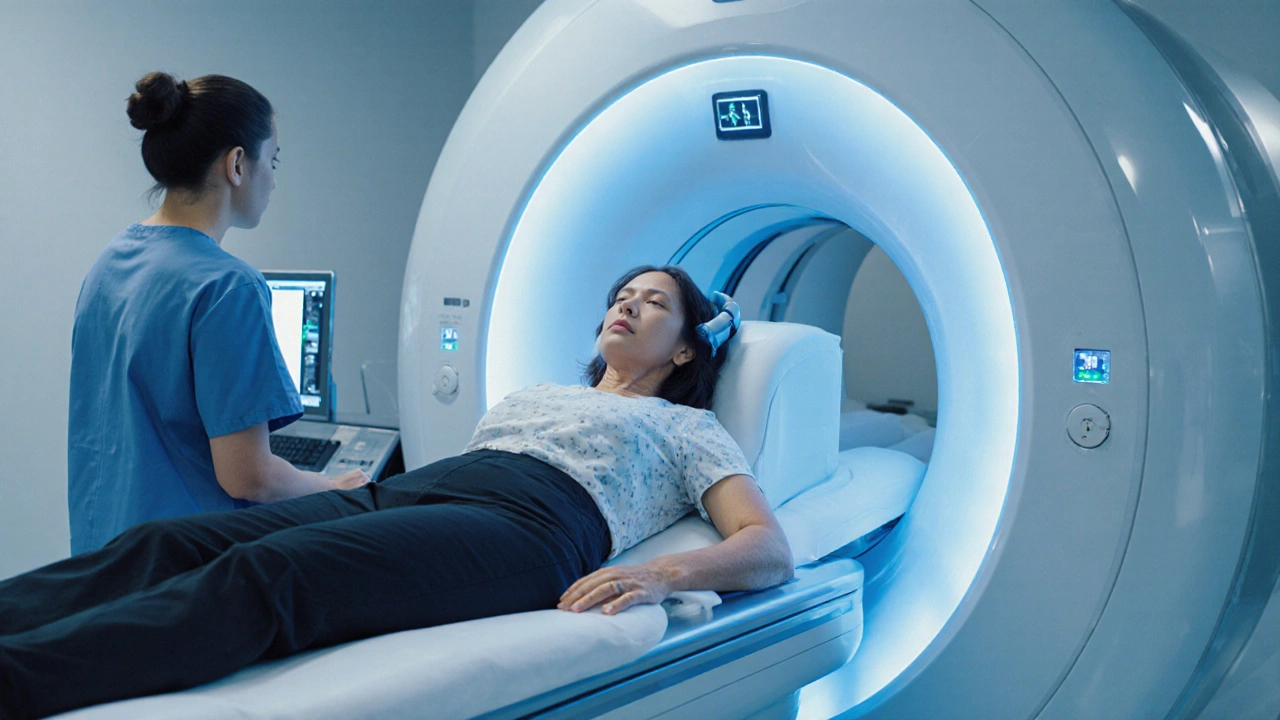
When a patient experiences Clinically Isolated Syndrome, a single neurological episode lasting at least 24hours that suggests demyelination and may be the first sign of multiple sclerosis, doctors turn to imaging for answers.
CIS can present as optic neuritis, a brainstem event, a spinal cord episode, or any focal neurological deficit. Roughly 30% of people with CIS will develop full‑blown multiple sclerosis within five years, making early, accurate diagnosis critical.
Why MRI Matters for CIS
Magnetic Resonance Imaging (MRI) provides unparalleled detail of soft tissue, allowing clinicians to spot the tiny white‑matter lesions that define demyelinating disease. Unlike CT, MRI does not use ionising radiation and can be repeated safely for monitoring.
Multiple Sclerosisa chronic immune‑mediated disorder characterized by demyelination in the central nervous system often begins with CIS, so the ability to visualize lesion load and location directly influences prognosis and treatment decisions.
Standard MRI Protocols for CIS
Guidelines from the 2024 International MS Consortium recommend a minimum protocol that covers both brain and spinal cord. The typical sequence set includes:
- T1‑weighted images (pre‑contrast) - provides anatomy and helps assess black holes.
- T2‑weighted images - highlights hyperintense lesions.
- Fluid‑attenuated inversion recovery (FLAIR) - suppresses CSF signal to reveal periventricular plaques.
- Gadolinium‑enhanced T1 - identifies active inflammation.
- Contrast agent: Gadolinium contrasta paramagnetic substance that shortens T1 relaxation, making active lesions appear bright.
- Spinal cord sagittal and axial T2 - catches cervical and thoracic plaques that may be missed on brain scans.
High‑resolution 3‑Tesla scanners are now the norm, but a well‑executed 1.5‑Tesla study still meets diagnostic criteria.
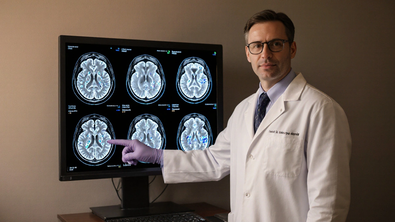
Typical MRI Findings in CIS
The 2025 McDonald criteria focus on three imaging hallmarks:
- Ovoid periventricular lesions - often described as “Dawson’s fingers” extending outward from the ventricles.
- Juxtacortical or cortical lesions - appear on FLAIR as bright spots adjacent to the grey‑matter surface.
- Infratentorial lesions - located in the brainstem or cerebellum, best seen on T2 and FLAIR.
When a spinal cord lesion is present, it typically spans less than two vertebral segments and is hyperintense on T2.
Lesion count matters: three or more brain lesions that meet the spatial criteria increase the odds of converting to multiple sclerosis by more than 70% within two years.
Interpreting MRI Results: Differentiating CIS From Mimics
Not every white‑matter bright spot means CIS. Common look‑alikes include small vessel ischemic disease, migraine‑related changes, and leukodystrophies. Radiologists use a combination of pattern, size, and location to rule these out.
Key discriminators:
- Age‑related white matter disease usually presents as diffuse, ill‑defined hyperintensities sparing the periventricular zone.
- Migraine lesions are often subcortical and asymmetrical, rarely enhancing with contrast.
- Infectious or inflammatory disorders such as neuromyelitis optica show longitudinally extensive spinal lesions >3 vertebral segments.
When doubt remains, a follow‑up MRI after 3-6months can reveal new lesions or enhancement, clarifying the diagnosis.
Next Steps After the Scan
Once MRI confirms the presence of demyelinating lesions, the clinical pathway typically includes:
- Referral to a neurologist specializing in demyelinating disease.
- Baseline neurological exam and visual evoked potentials.
- Discussion of disease‑modifying therapies if the risk of conversion to multiple sclerosis is high.
- Schedule a repeat MRI at 3‑month intervals during the first year to monitor lesion activity.
Patients without any lesions on MRI are considered low‑risk, but they still need symptom‑focused care and education about warning signs.
Common Pitfalls & Pro Tips
- Pitfall: Skipping spinal cord imaging can miss crucial lesions. Pro tip: Always include a cervical and thoracic T2 sequence.
- Pitfall: Using only T1 without contrast may underestimate active disease. Pro tip: Add gadolinium‑enhanced T1 when the initial scan is inconclusive.
- Pitfall: Over‑relying on a single MRI for prognosis. Pro tip: Combine imaging with clinical and cerebrospinal fluid markers for a balanced view.
Table: MRI Sequences vs Diagnostic Value for CIS
| Sequence | Primary Insight | Strengths | Limitations |
|---|---|---|---|
| T2‑weighted | Lesion detection | High sensitivity for white‑matter plaques | Cannot differentiate active vs chronic lesions |
| FLAIR | Periventricular & cortical lesions | Suppresses CSF, reveals Dawson’s fingers | Longer acquisition time |
| Gadolinium‑enhanced T1 | Active inflammation | Shows blood‑brain barrier breakdown | Requires IV contrast, may miss non‑enhancing lesions |
| Spinal cord T2 | Infratentorial disease | Detects cervical/thoracic plaques | Susceptible to motion artefacts |

Frequently Asked Questions
Can a normal MRI rule out CIS?
A normal MRI makes CIS unlikely but does not guarantee the absence of demyelination. Small lesions below the resolution of standard scanners or purely clinical episodes can still qualify as CIS. Follow‑up imaging is advised if symptoms persist.
How soon after symptom onset should the MRI be performed?
Ideally within 2‑4weeks. Early imaging captures active enhancement, which may fade after a month, potentially missing critical diagnostic information.
Is gadolinium contrast safe for repeated use?
Current evidence indicates that standard doses are safe for most adults, but patients with severe kidney disease should avoid it. Recent guidelines suggest limiting contrast‑enhanced scans to situations where they directly affect management.
What MRI findings predict conversion to multiple sclerosis?
Three or more brain lesions that meet the spatial criteria (periventricular, juxtacortical, or infratentorial) plus at least one gadolinium‑enhancing lesion increase conversion risk to over 70% within two years.
Do spinal cord lesions affect prognosis?
Yes. The presence of cervical spinal cord lesions, especially those that are T2‑hyperintense and less than two vertebral segments long, is associated with a higher likelihood of early conversion to multiple sclerosis.

Ashley Allen
September 29, 2025 at 23:23Thanks for the thorough overview of the MRI protocol. The checklist will be handy when discussing CIS with patients.
Brufsky Oxford
October 3, 2025 at 10:43The transition from a single clinical event to a chronic demyelinating disease invites a deep reflection on how we define pathology.
In the early days of neuroimaging, the magnetic resonance scanner was merely a novelty, yet it quickly became the oracle for neurologists.
Today, the MRI not only confirms the presence of lesions but also maps the temporal landscape of inflammation.
When a patient presents with optic neuritis, the urgency to capture gadolinium enhancement within weeks is a race against the invisible fire of immune attack.
The periventricular “Dawson’s fingers” are more than radiological curiosities; they are anatomical testimonies of immune cells navigating the venous architecture.
Likewise, spinal cord lesions, though often small, betray a propensity for early clinical conversion.
The McDonald criteria, refined over decades, embed these imaging hallmarks into a probabilistic framework that guides therapeutic decisions.
Yet one must remember that imaging is a snapshot, a momentary still of a dynamic process.
Serial scans at three‑month intervals transform that snapshot into a motion picture, revealing new plaques or resolving inflammation.
In this context, the risk calculator becomes a conversational bridge, translating raw lesion counts into patient‑centred prognostic language.
The calculator’s thresholds-two lesions versus three active lesions-mirror the underlying biology of lesion dissemination.
A high‑risk assessment (>70% conversion) should prompt clinicians to discuss disease‑modifying therapies with candor and empathy.
Conversely, a low‑risk profile offers an opportunity for shared decision‑making about monitoring versus early intervention.
It is also worth noting that some white‑matter hyperintensities arise from vascular etiologies, underscoring the need for clinical correlation.
Ultimately, the MRI serves as both a diagnostic lighthouse and a prognostic compass, steering patients through uncertain seas.
So, while the technology is impressive, the real art lies in integrating these images with the patient’s story and goals :).
VAISHAKH Chandran
October 6, 2025 at 22:03One must appreciate that the sophisticated nuances of neuro‑imaging are lost on the lay public who skim headlines and think a lesion is a mere artifact the radiologist can overlook yet the reality is that without the proper high‑field strength protocol the subtleties of periventricular demyelination remain invisible.
Pat Merrill
October 10, 2025 at 09:23Oh sure, because we all have 3‑Tesla scanners in every clinic and the budget for gadolinium is endless, apparently. It's definitely not like many community hospitals struggle to get even a basic T2‑FLAIR done. The whole pretentious elitist vibe is pretty funny when you consider that most patients just want to know if their vision will improve, not a PhD dissertation on imaging physics.
Vicki Roth
October 13, 2025 at 20:43The inclusion of spinal cord imaging is essential for accurate CIS assessment.
Vishal Bhosale
October 17, 2025 at 08:03Spinal MRI is often ignored but it holds key info the brain scan alone can't provide and missing it can lead to underestimating risk.
Garima Gauttam
October 20, 2025 at 19:23While the risk calculator tries to quantify uncertainty it inevitably reduces a complex neuro‑immune process to a tidy number which many clinicians find comforting yet misleading.
Georgia Nightingale
October 24, 2025 at 06:43It's true that any algorithm simplifies reality, however the McDonald criteria themselves are built on decades of empirical observation and have stood the test of clinical trials. The calculator simply operationalizes those criteria, turning lesion count and enhancement into actionable risk percentages. When used appropriately it enhances shared decision‑making rather than replacing nuanced clinical judgment. In practice, doctors who understand the limitations can still rely on the tool to guide discussions about early treatment initiation.
Chris Kivel
October 27, 2025 at 17:03Great summary of the MRI sequences; the table makes it easy to remember which protocol adds the most diagnostic value.
Fredric Chia
October 31, 2025 at 04:23While the table is concise, it overlooks the impact of motion artefacts on spinal imaging, a flaw that undermines its utility.
Julia Odom
November 3, 2025 at 15:43Seeing the clear breakdown of each sequence really empowers clinicians to approach CIS evaluation with confidence. The vivid description of Dawson's fingers and the emphasis on early gadolinium use serve as actionable reminders. By integrating these protocols into routine practice we not only improve diagnostic accuracy but also foster timely therapeutic decisions that can alter disease trajectory. This kind of structured guidance bridges the gap between research and bedside care, ultimately benefiting patients.
Danielle Knox
November 7, 2025 at 03:03Sure, because a tidy table and fancy adjectives are all it takes to change a patient’s outcome, right? In reality the real work happens in the follow‑up and adherence, not just in the pretty presentation.