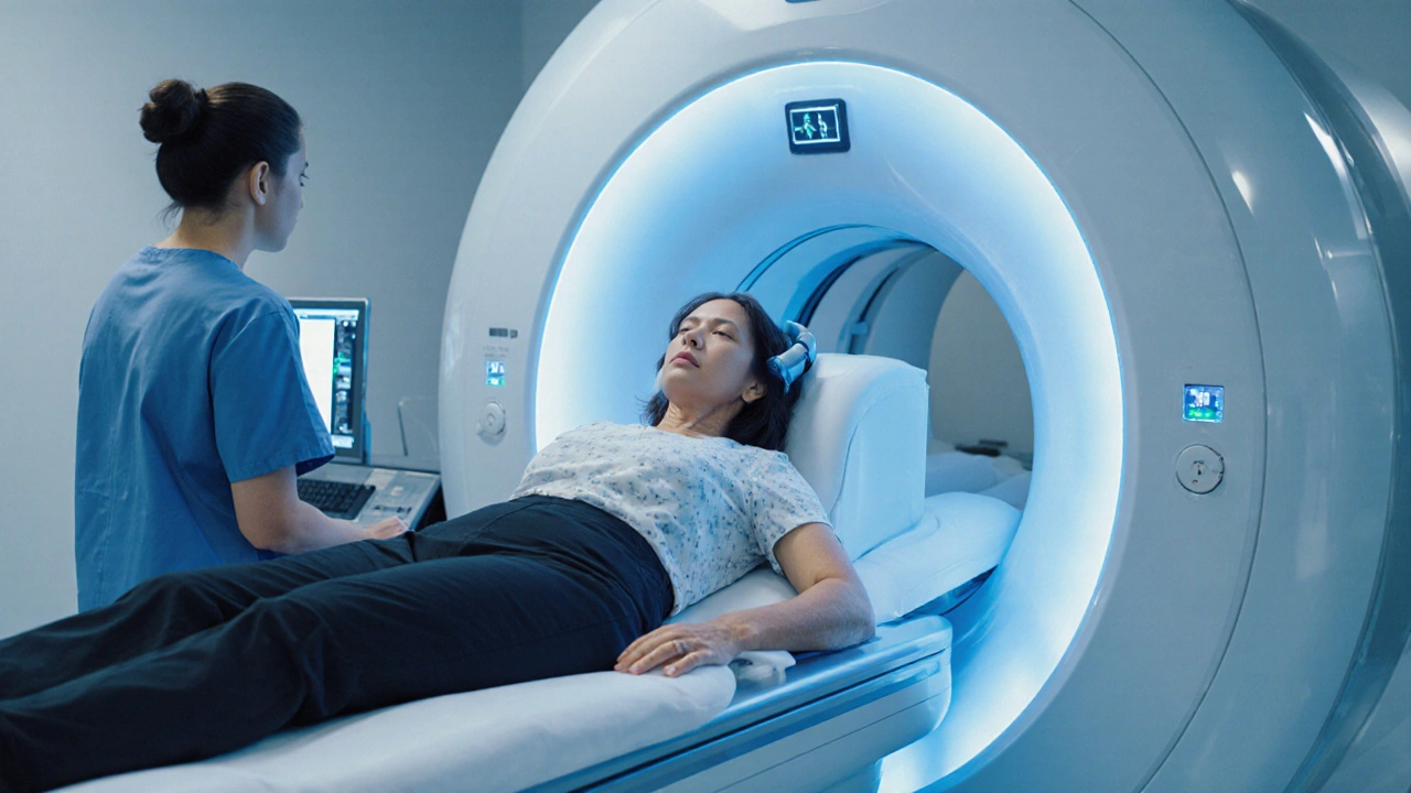
Explore how MRI works to diagnose Clinically Isolated Syndrome, the key imaging protocols, typical findings, and next steps for patients and clinicians.
Clinically Isolated Syndrome – Overview
When working with Clinically Isolated Syndrome, a first‑time neurological event that appears as a single demyelinating lesion on brain or spinal‑cord imaging. Also known as CIS, it signals a possible early stage of a larger autoimmune disorder and warrants careful monitoring. In everyday language, think of it as the body’s first warning flag that the myelin sheath around nerves might be under attack. The condition Clinically Isolated Syndrome encompasses a single demyelinating episode, and physicians usually ask three critical questions: what symptoms appeared, where the lesion is located, and whether any other invisible lesions show up on scans. These answers shape the next steps—whether to watch, test further, or start treatment. Because CIS can be the first chapter of a longer story, early detection matters: catching it before it spreads can change a patient’s long‑term outlook.
Key Players Around Clinically Isolated Syndrome
One of the biggest names linked to CIS is Multiple Sclerosis, a chronic, immune‑mediated disease that creates multiple demyelinating patches throughout the central nervous system. Research shows that up to 30 % of people with CIS eventually meet the diagnostic criteria for Multiple Sclerosis, so clinicians treat CIS as a possible gateway. Another central tool is MRI, magnetic resonance imaging that visualizes brain and spinal‑cord lesions with high detail. MRI influences early detection of CIS by revealing hidden lesions that a physical exam alone would miss, and the number of these silent lesions helps estimate conversion risk to Multiple Sclerosis. Beyond these, the autoimmune demyelination process itself acts as the underlying mechanism, meaning the body’s own immune cells mistakenly attack myelin. Diagnosis criteria—often referred to as the 2017 McDonald criteria—require a combination of clinical evidence, MRI findings, and sometimes cerebrospinal‑fluid analysis. These criteria create a clear roadmap: if MRI shows at least one additional lesion in a characteristic location, the probability of progression rises sharply. Understanding how these entities interact—CIS, MRI, Multiple Sclerosis, and the diagnostic framework—gives patients and doctors a shared language for decision‑making.
Armed with these basics, you’ll find a curated set of articles below that dive deeper into treatment options, lifestyle tweaks, and emerging research tied to Clinically Isolated Syndrome. Whether you’re a patient curious about next‑step options, a caregiver looking for practical advice, or a clinician seeking the latest evidence, the collection covers everything from risk assessment to therapy choices. Browse the posts to see how experts translate this complex web of entities into real‑world actions you can take today.

