Explore how MRI works to diagnose Clinically Isolated Syndrome, the key imaging protocols, typical findings, and next steps for patients and clinicians.
Brain Imaging: Tools, Techniques, and Clinical Insights
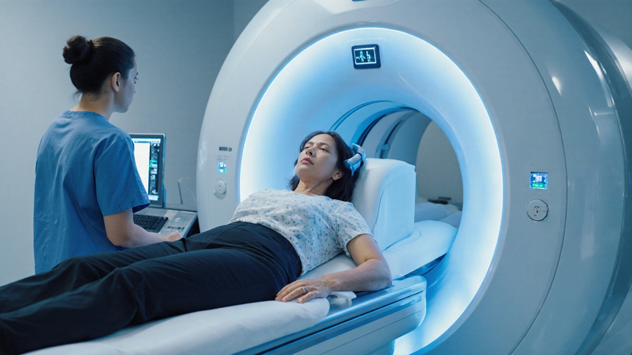
When exploring brain imaging, the set of medical technologies that create pictures of the brain’s structure and function. Also known as neuroimaging, it helps doctors see what’s happening inside the skull without surgery. One of the most common methods is MRI, magnetic resonance imaging that uses powerful magnets and radio waves to generate detailed, high‑resolution images of brain tissue, often called the gold standard for soft‑tissue detail. CT scan, computed tomography that employs X‑rays to produce cross‑sectional slices, excels at spotting acute bleeding and bone injuries, while PET, positron emission tomography that tracks metabolic activity using radioactive tracers, reveals functional changes linked to Alzheimer’s or epilepsy. Together these tools **brain imaging** encompasses structural, vascular, and metabolic assessment, requires specialized equipment and trained radiologists, and influences treatment plans across neurology, oncology, and psychiatry.
How Different Modalities Fit Into Real‑World Care
Understanding each modality’s strengths lets clinicians match the right test to the patient’s problem. MRI shines when doctors need to examine tumors, demyelinating disease, or subtle inflammation because its contrast resolution far exceeds CT. For emergency settings, a CT scan can be performed in minutes, making it the first line for suspected stroke, head trauma, or intracranial hemorrhage. PET adds a functional layer, showing where glucose metabolism is reduced in early Alzheimer’s or how a brain tumor responds to chemotherapy. Advanced techniques like functional MRI (fMRI) map brain activity during tasks, guiding neurosurgeons to avoid critical regions during tumor removal. Contrast agents—gadolinium for MRI, iodine‑based for CT—enhance vascular detail, but they also require safety checks for kidney function. Radiologists interpret these images alongside clinical data, often collaborating with neurologists, oncologists, and surgeons to decide on medication choices such as antiepileptics, chemotherapy agents, or disease‑modifying drugs. This multidisciplinary loop mirrors many of the medication guides on our site, where accurate imaging informs the selection of treatments like pirfenidone for fibrotic diseases or targeted therapies for cancers.
Whether you’re a patient curious about why a doctor ordered an MRI, a caregiver planning the next steps after a CT scan, or a health professional needing a refresher on PET interpretation, the articles below break down each technology, explain preparation tips, and highlight what findings mean for treatment decisions. You’ll find practical advice on scheduling scans, understanding reports, and navigating insurance, plus deeper dives into how imaging shapes drug choices for conditions ranging from oral cancer to HIV. Dive into the collection to see real‑world examples, safety considerations, and actionable takeaways that will help you make sense of every brain imaging result you encounter.